AI Cytology Cuts Time to Detect Dog Cancer
Veterinary Practice News reports that AI-supported digital cytology can provide cytology results in about 20 minutes, enabling same-visit diagnosis and treatment planning for canine masses.
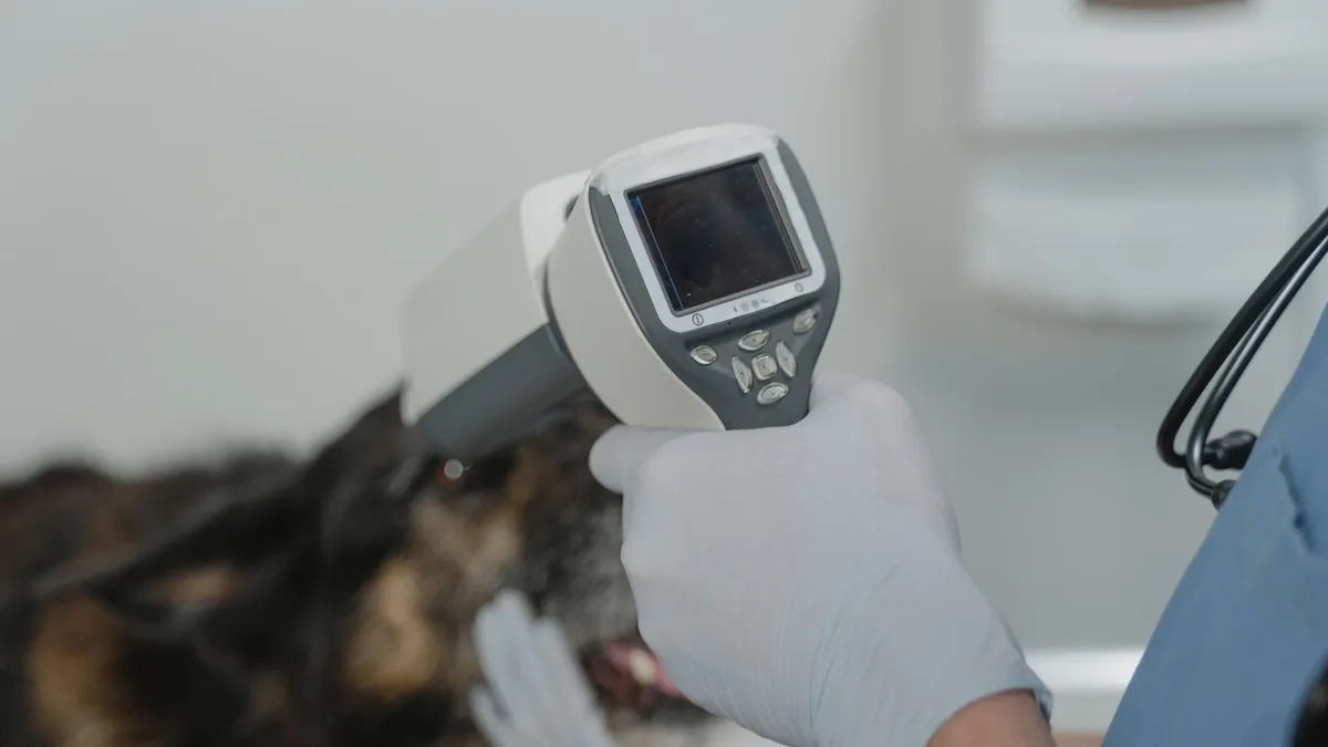
Veterinary Practice News reports that AI-supported digital cytology can deliver results in about 20 minutes, allowing veterinarians to discuss diagnosis and treatment during the same visit. Recent research reports that 21 percent of pet owners found the death of their pet the most distressing loss they had ever experienced.
The technology is part of the See Something, Do Something. Why Wait? Aspirate.® program, which encourages teams to aspirate masses the size of a pea (1 cm) that have been present for one month, as outlined by the squad of veterinary professionals.
Fine-needle aspiration (FNA) is a minimally invasive technique that can distinguish inflammation, benign growth, or malignancy without rushing the patient into surgery.
However, sample quality and interpretation can vary, and conventional external laboratories add cost and delay, sometimes several days, for owners who may already be anxious.
In the U.S., nearly 40 percent of veterinary professionals now incorporate AI into their everyday practice, according to recent research, as shown in the stats of veterinary practice.
AI-supported digital cytology scans the slide and can provide useful information quickly, often in the same visit, with results typically available in about 20 minutes.
When the report indicates a mast cell tumor (MCT), the veterinarian can explain treatment options immediately, such as staging, surgery, intratumoral injection, referral, or expert review.
The AI report still requires veterinary judgment; it must be interpreted in the context of the patient, physical exam, sample quality, and what was seen on the slide.
In complex cases, a clinical pathologist’s expert review can add value, especially for cytologic grading of MCTs, which informs staging and treatment recommendations.
The value of AI-supported digital cytology lies in its practicality, efficiency, and cost-effectiveness as a screening tool, helping identify benign processes and diagnosing malignant MCTs, the most common malignant cutaneous cancer in dogs, as demonstrated in recent fixtures of diagnostic workflows.
Case in context: Zoe, an eight-year-old spayed female American bulldog, presented for a recheck of follicular lymphoma with salivary gland involvement.
During the exam, a new flat, movable subcutaneous mass was noted on her left ventrolateral abdomen.
The FNA was performed and submitted through AI-supported digital cytology, which identified findings consistent with an MCT.
Having the information available during the visit changed the conversation; the owner did not have to wait days for results.
Based on the AI findings, an add-on expert review confirmed a low-grade MCT, and surgical removal was recommended.
Zoe underwent surgery eight days later; histopathology confirmed a low-grade subcutaneous MCT with no infiltrative pattern, no multinucleated cells, and a mitotic count of 0.
Margins were narrow laterally but complete, with a good deep margin and a tissue plane deep to the tumor.
A separate ventral thoracic lipoma was also removed, and the owner returned for a post-operative oncology recheck to review the biopsy report in detail.