Study Compares Cytology and Histopathology for Canine Splenic Masses
A veterinary study finds significant diagnostic disagreement between cytology and histopathology for canine splenic nodules.
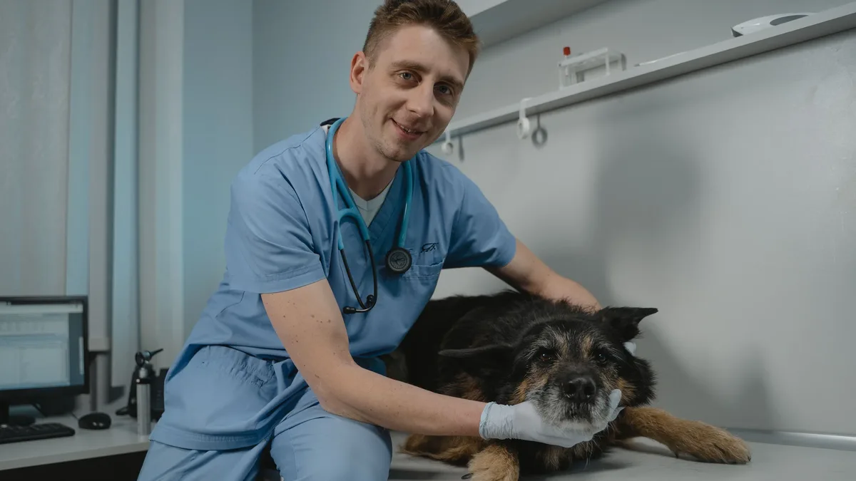
New research examines the reliability of cytology for diagnosing canine splenic masses, revealing notable discrepancies with surgical pathology. A separate study of diagnostic methods finds a modern PCR panel identifies parasites in pet stool samples far more frequently than conventional tests.
Cytology Accuracy for Splenic Masses
A recent study published in JAVMA evaluated the correlation between cytology and histopathology results for splenic masses in dogs. The research, led by University of Georgia professor of small animal surgery Janet Grimes, DVM, MS, DACVS, retrospectively reviewed records from 33 dogs that had both cytologic and histopathologic evaluation of a splenic mass or nodule within 90 days. Agreement between ultrasound-guided fine-needle aspirate cytology and histopathology from splenectomy or necropsy on a diagnosis of neoplastic versus nonneoplastic conditions ranged from 55% to 67%, with disagreement occurring in 24% of cases.
The study advises that cytology cannot rule out a neoplastic condition but can suggest one, thereby endorsing histopathology for a definitive diagnosis. For dogs with smaller, incidentally identified splenic nodules, Dr. Grimes notes that cytology can guide clinical decision-making. If cytology indicates neoplasia, splenectomy is recommended. However, a non-neoplastic cytology result does not fully rule out underlying malignancy, necessitating active surveillance with abdominal ultrasound every three months. Dr. Grimes stated, "Active surveillance should include an abdominal ultrasound every 3 months and decisions made on new findings." The study's limitations include its focus on non-ruptured lesions and the potential difficulty in matching aspirated nodules to corresponding histopathology slides in dogs with multiple masses.
Fecal PCR Panel Efficacy
A fecal quantitative polymerase chain reaction (qPCR) panel detected parasites in twice as many canine and feline fecal samples as antigen testing and centrifugal flotation combined. The study, published in Parasites & Vectors, analyzed 1,013 stool samples (882 from dogs, 131 from cats) collected from 19 states in May and June 2025. Researchers compared the KeyScreen GI Parasite PCR panel from Antech Diagnostics with the Fecal Dx Antigen Profile with Giardia and zinc sulfate centrifugal flotation performed at IDEXX Laboratories.
The qPCR panel, which targets 20 parasites, detected at least one parasite in 27.2% of samples. In contrast, antigen testing found parasites in 10.9% of samples, and flotation detected them in 6.5%. Together, the two conventional methods identified parasites in 13.6% of samples. Overall, 70.9% of samples were negative on all three methods, while 15.5% were positive only on the qPCR panel.
The panel's advantage varied significantly by parasite type. Its detection rates were higher for Giardia duodenalis, flea tapeworm (Dipylidium caninum), and Cystoisospora species. Whipworm detection was also higher with qPCR, though not statistically significant. Roundworm detection was comparable between methods. Interestingly, the hookworm antigen test detected more positives than the qPCR panel, a difference attributed to proteins secreted by adult hookworms before egg production. The qPCR panel also detected protozoa not targeted by the antigen panel, including Tritrichomonas blagburni and Cryptosporidium species. Eimeria species were the most common finding among the qPCR-only detections.